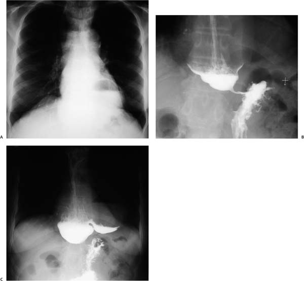
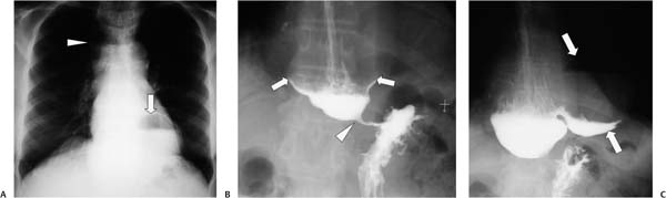
Case 100 A 71-year-old man presents with halitosis. (A) Frontal chest radiograph shows a large, air-filled structure extending from the upper to the lower chest (arrowhead) and a large air-fluid level in the retrocardiac left lower chest (arrow). (B) Barium study shows a markedly dilated esophagus, or “megaesophagus” (arrows), a “rat tail” deformity of the lower esophageal sphincter (arrowhead), and slow passage of contrast into the stomach. (C) Delayed image shows a large, round paraesophageal collection containing an air-fluid level and dependent layering of barium (arrows). • Achalasia with epiphrenic diverticulum: This is the top choice, given the megaesophagus, the rat tail stricture of the distal esophagus, and an epiphrenic saccular outpouching from the esophagus. • Secondary achalasia and pseudoachalasia:
 Clinical Presentation
 Imaging Findings
 Differential Diagnosis
![]()
Stay updated, free articles. Join our Telegram channel

Full access? Get Clinical Tree


