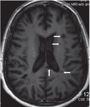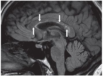

FINDINGS Figure 101-1. Axial FLAIR through the corona radiata. There are multifocal hyperintense lesions of variable shapes and sizes in the periventricular, deep, and subcortical white matter (WM) in the bilateral corona radiata (transverse arrows), some smudgy hyperintensities around the occipital horns. There is a left splenium callosal–septal interface lesion (vertical arrow). Figure 101-2. Axial T1WI through same level as in Figure 101-1. Some lesions show black holes (arrows). Figure 101-3. Sagittal T2 FLAIR. There are multiple callosal–septal interface hyperintense lesions (arrows).
DIFFERENTIAL DIAGNOSIS Toxic metabolic encephalopathy (medication induced, cocaine, inhaled opiates, chemoradiation, Marchiafava-Bignami), inflammatory demyelination (acute disseminated encephalomyelitis [ADEM] and Neuromyelitis optica [NMO]), systemic lupus erythematosus (SLE) Susac syndrome (SS) Multiple sclerosis (MS).
DIAGNOSIS Multiple sclerosis (MS).
DISCUSSION
Stay updated, free articles. Join our Telegram channel

Full access? Get Clinical Tree


