Case 102
Case History
A 39-year-old woman presents with a right breast palpable lump. She is 6 months post-partum and is still breast-feeding. She was initially studied with lower-frequency sonography (Fig. 102–2). Her sonogram was interpreted as BI-RADS Category 3 (probably benign lesion, short-term follow-up recommended). After 1 month, because symptoms persisted, she was examined with high-frequency sonography (Fig. 102–3). As a result of her high-frequency sonogram, mammograms were performed (Fig. 102–1).
Physical Examination
• right breast: palpable lump at the 6:00 position
• left breast: normal exam
Mammogram
Calcifications (Fig. 102–1)
• type: pleomorphic/heterogeneous
• distribution: grouped/clustered
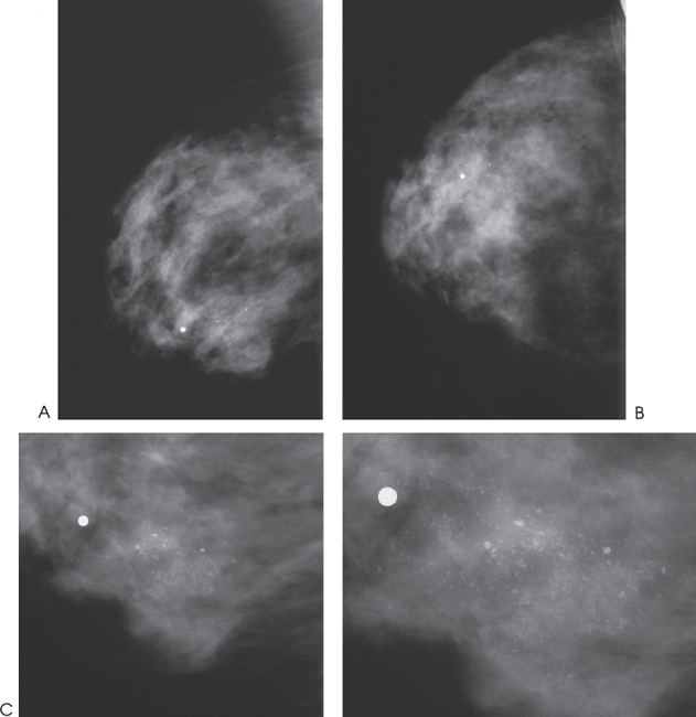
Figure 102–1. Near the right 6:00 position, there is a cluster of heterogeneous calcifications that corresponds to a palpable lump, which is marked with a radiopaque marker. (A). Right MLO mammogram. (B). Right CC mammogram. (C). Right MLO magnification mammogram.
Ultrasound
Low Frequency
Frequency
• 7 MHz
Mass
• margin: well defined, lobulated
• echogenicity: hypoechoic
• retrotumoral acoustic appearance: bilateral edge shadowing


Stay updated, free articles. Join our Telegram channel

Full access? Get Clinical Tree


