Case 103
Case History
A 51-year-old woman presents with new left breast calcifications on her screening mammogram.
Physical Examination
• normal exam
Mammogram
Calcifications (Fig. 103–1)
• type: pleomorphic/heterogeneous
• distribution: grouped/clustered
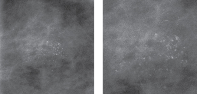
Figure 103–1. Left CC magnification mammogram: A cluster of heterogeneous calcifications is present in the outer breast.
Ultrasound
Frequency
• 10 MHz
Mass


Stay updated, free articles. Join our Telegram channel

Full access? Get Clinical Tree


