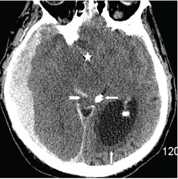
FINDINGS Figure 103-1. Axial NCCT through the tentorial incisura. There is a 1.5-cm thick crescentic hyperdense right temporal–occipital extraaxial collection displacing the right temporal and occipital lobes from the inner skull table consistent with acute subdural hematoma (ASDH) (transverse arrows). There is hyperdense subdural hematoma (SDH) around the right tentorium (triangle). There is crowding of the incisura with effacement of the cerebrospinal fluid (CSF) spaces. The right temporal horn is obliterated. The left temporal horn is severely dilated with periventricular hypodensity (vertical arrow) consistent with transependymal CSF permeation due to the acute hydrocephalus. Figure 103-2. Axial NCCT through the suprasellar cistern. There is obliteration of the suprasellar (star), interpeduncular, perimesencephalic, and quadrigeminal cisterns by a right uncal and transtentorial herniation. The left trigone is massively dilated (vertical arrow). There is acute hemorrhage (hyperdensity) in the right quadrigeminal cistern/midbrain (chevron). The calcified pineal gland is displaced inferiorly and to the left—midline shift (transverse arrow).
DIFFERENTIAL DIAGNOSIS N/A.
DIAGNOSIS Acute subdural hematoma (ASDH) with complications.
DISCUSSION
Stay updated, free articles. Join our Telegram channel

Full access? Get Clinical Tree


