Case 104
Case History
A 72-year-old woman presents with new calcifications on her right screening mammogram.
Physical Examination
• right breast: two palpable cysts at the 10:00 and 6:00 position; a third palpable nodule adjacent to the cyst at 10:00, corresponding to a solid mass
• left breast: one palpable cyst
Mammogram
Calcifications (Figs. 104–1 and 104–2)
• type: pleomorphic/heterogeneous
• distribution: grouped/clustered
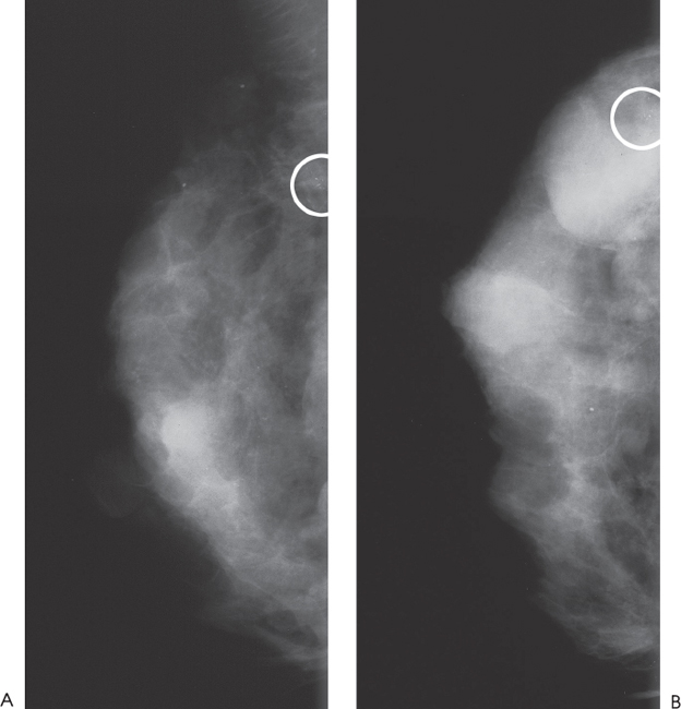
Figure 104–1. The breast is extremely dense and contains multiple round masses that sonographically correspond to cysts. In the upper outer breast there is a cluster of calcifications (circled), which corresponds to a sonographically solid mass. (A). Right MLO mammogram. (B). Right CC mammogram.


Stay updated, free articles. Join our Telegram channel

Full access? Get Clinical Tree


