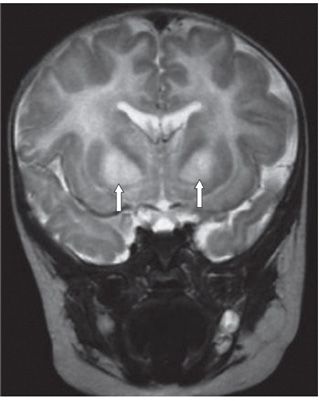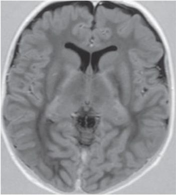

FINDINGS Figure 104-1. Axial T2WI through the basal ganglia. There is diffuse white matter (WM) hyperintensity with involvement of the globus pallidus and anterior aspect of the cerebral peduncles (arrows). Figure 104-2. Coronal T2WI through the basal ganglia. There is hyperintensity in the WM and bilateral globus pallidus (arrows). Figure 104-3. Axial T1 inversion recovery image through the basal ganglia. There is marked hypointensity throughout the WM.
DIFFERENTIAL DIAGNOSIS Pelizaeus-Merzbacher disease, Alexander disease, Canavan disease mucopolysaccharidoses, megalencephalic leukodystrophy with cysts.
DIAGNOSIS Canavan disease.
DISCUSSION
Stay updated, free articles. Join our Telegram channel

Full access? Get Clinical Tree


