FINDINGS All MRV images are contrast-enhanced brain MRV, MIP, or volume rendered in appropriate projections to show the sinuses.
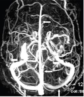
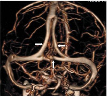
Figure 105-1. Sagittal MIP. The superior sagittal sinus (SSS) (vertical arrows) drains the cortical veins into the torcula (star). The largest of the cortical veins is the vein of Trolard or the superior anastomotic vein (transverse arrows) connecting the SSS to the middle cerebral vein of Sylvius. The deep structures are drained by the paired internal cerebral veins (ICVs) (line arrows). The ICVs are joined by the basal veins of Rosenthal (pentagon) to form the great vein of Galen (GVG) (arrowhead). The GVG is connected to the torcula by the straight sinus (SS) (chevron). On this sagittal MIP, the bilateral transverse and sigmoid sinuses are superimposed in the posterior fossa (curved arrow). The occipital sinus is a midline structure (triangle) draining into the torcula. Figure 105-2


Stay updated, free articles. Join our Telegram channel

Full access? Get Clinical Tree


