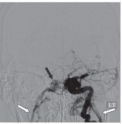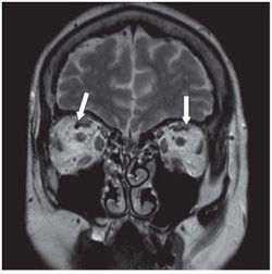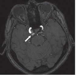


FINDINGS Figure 106-1. Coronal NCCT through the orbits. There is enlargement of the left superior ophthalmic vein relative to the right (arrows). Figure 106-2. DSA left internal carotid artery (ICA) PA (posterior-anterior) view. There is abnormal early and dense opacification of the left cavernous sinus with early opacification of the contralateral right cavernous sinus (black arrow) and the bilateral jugular veins (white arrows). Figure 106-3. Coronal T2WI through the orbits in a companion case with incidental findings at imaging for unrelated complaint. There is mild prominence of the right superior ophthalmic vein (arrows). Figure 106-4. Time of flight MRA source images level of the cavernous sinus in the same patient as figure 106-3. There is asymmetric flow-related signal in the right cavernous sinus.
DIFFERENTIAL DIAGNOSIS Cavernous sinus thrombosis, carotid cavernous fistula (CCF), cavernous ICA aneurysm.
DIAGNOSIS Both patients have CCF.
Stay updated, free articles. Join our Telegram channel

Full access? Get Clinical Tree


