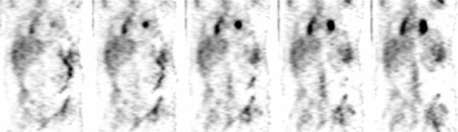
CASE 106 A 48-year-old man with uveitis is referred by ophthalmology for suspected sarcoidosis. Fig. 106.1 Whole-body planar image, anterior and posterior projections, 67Ga. Fig. 106.2 Selected “whole-body” SPECT image, coronal projection, 67Ga. • A 7.5 mCi dose of 67Ga-citrate is injected intravenously 3 days before scan. • Whole-body imaging in anterior and posterior projections Whole-body planar images (Fig. 106.1) and “whole-body” SPECT images (Fig. 106.2) demonstrate intense right paratracheal and symmetric bilateral hilar uptake; this pattern has been termed the lambda sign because it resembles the Greek letter. Coupled with intense, symmetric uptake in the parotid glands and lacrimal glands, this is a characteristic pattern of sarcoidosis. The colon activity is physiologic. • Sarcoidosis • Lymphoma • Inflammatory/infectious lymphadenitis Active sarcoidosis. No follow-up obtained.
Clinical Presentation
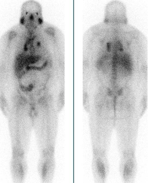
Technique
 Dual-detector gamma camera
 Medium-energy collimators
 Energy peak at 93-, 185-, and 300-keV photopeaks
Image Interpretation
Differential Diagnosis
Diagnosis and Clinical Follow-Up
Discussion
Related posts:


Stay updated, free articles. Join our Telegram channel

Full access? Get Clinical Tree


