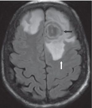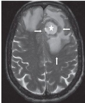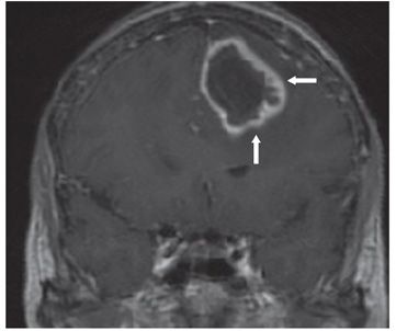


FINDINGS Figure 107-1. Axial DWI MRI through the frontal lobes. There is a left frontal lobe well-circumscribed hyperintense mass (arrows) consistent with restricted diffusion. A similar but smaller lesion is present inferiorly in the right frontal lobe (not shown). Figure 107-2. Axial FLAIR through the same level. There is alternating (six) layers of different intensity pattern (concentric target pattern) (transverse arrows) within the left frontal lobe lesion. There is surrounding confluent hyperintensity consistent with vasogenic edema (vertical arrow) and mass effect on surrounding structures manifested by effacement of convexity sulci. There is vasogenic edema in the right frontal lobe surrounding the second lesion (not shown). Figure 107-3. Axial T2WI through the left frontal lobe mass. There is a central high intensity core (star) with a surrounding multi-layer irregular wall; isointense to brain around the core with an outer layer of thin hyperintensity (transverse arrows) before the edema (vertical arrow). Figure 107-4. Post-contrast coronal T1WI through the left frontal mass. There is a thick irregular mural contrast enhancement (arrow). Axial non-contrast T1WI (not shown) demonstrated a hypointense mass surrounded by a thin mildly hyperintense wall and another coat of isointense capsule before the vasogenic edema.
DIFFERENTIAL DIAGNOSIS Abscess (pyogenic), toxoplasmosis, aspergillosis, glioblastoma (GB), infarcts.
DIAGNOSIS
Stay updated, free articles. Join our Telegram channel

Full access? Get Clinical Tree


