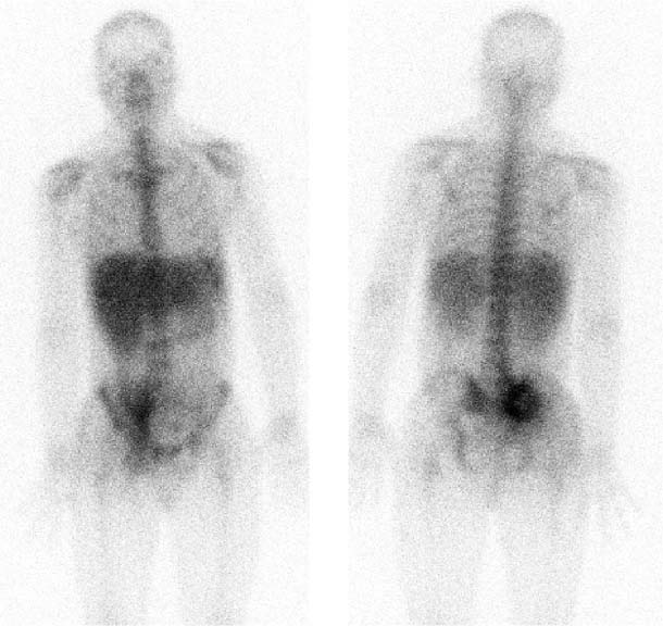
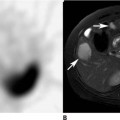
CASE 107 A 36-year-old man with a history of intravenous drug abuse presents with right hip and flank pain radiating down his right leg. Fig. 107.1 Whole-body planar image, anterior and posterior projections, 67Ga. • A 7.5 mCi dose of 67Ga-citrate is injected intravenously 3 days before scan. • Whole-body imaging in anterior and posterior projections Intense tracer uptake is noted in the region of the right sacroiliac joint (Fig. 107.1). Note the “cold” center on the posterior view; this suggests an advanced process with central devitalization (necrosis). Faint tracer uptake is apparent in the soft tissues of the right buttock. • Osteomyelitis • Sacroiliitis • Soft tissue inflammation/infection • Acute trauma • Malignancy Findings consistent with unilateral sacroiliitis (with osteomyelitis) and adjacent cellulitis. Correlative MRI demonstrates local sacroiliac bone destruction and edema of adjacent gluteus muscles.
Clinical Presentation
Technique
 Dual-detector gamma camera
 Medium-energy collimators
 Energy peak at 93-, 185-, and 300-keV photopeaks
Image Interpretation
Differential Diagnosis
Diagnosis and Clinical Follow-Up
Discussion
Related posts:


Stay updated, free articles. Join our Telegram channel

Full access? Get Clinical Tree


