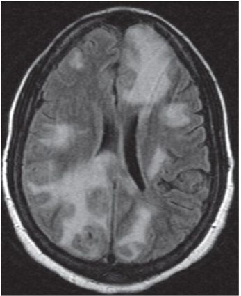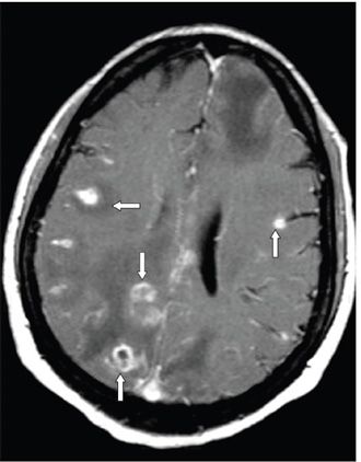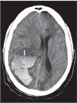


FINDINGS Figure 108-1. Axial NCCT through the basal ganglia. There are multiple patchy hypodense areas in the white matter (WM) (arrows). Figure 108-2. Axial FLAIR through the corona radiata. There are large hyperintense areas in bilateral cerebral WM with local mass effect. Figure 108-3. Corresponding axial post-contrast T1WI. There are multiple areas of enhancement mostly nodular and ring with surrounding edema (arrows). Figure 108-4. Axial NCCT in a different patient shows a large right parieto-occipital acute hemorrhage with an internal fluid level (arrow) as a complication of intracerebral posttransplant lymphoproliferative disorder (PTLD).
DIFFERENTIAL DIAGNOSIS Abscess, toxoplasmosis, and other neoplastic processes (other central nervous system [CNS] lymphomas), posttransplant lymphoproliferative disorder.
Stay updated, free articles. Join our Telegram channel

Full access? Get Clinical Tree


