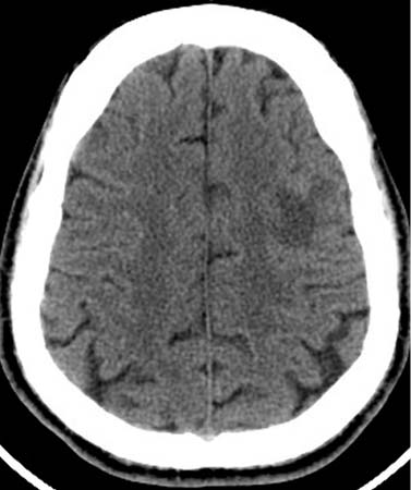
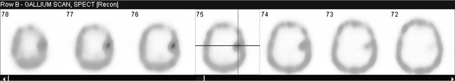
CASE 108 A 45-year-old man, a heroin user and HIV-positive, presents with headache and fever. CT of the head shows a low-attenuation lesion in the left frontal lobe (Fig. 108.1). Fig. 108.1 CT, axial image. Fig. 108.2 Sequential 201Tl SPECT, axial images. Fig. 108.367Ga SPECT. • A 4.0 mCi dose of 201Tl is injected intravenously for scan on day 1. • A 7.5 mCi dose of 67Ga is injected intravenously on day 1 (after 201Tl scan) for scan on day 4. • Sequential 201Tl SPECT imaging of the brain on day 1 with a dual-detector gamma camera CT scan reveals a mass lesion in the left frontal lobe of the brain. Normally, both 201Tl and 67Ga are excluded from the cerebrum. This lesion is thallium-negative (Fig. 108.2) but gallium-positive (Fig. 108.3
Clinical Presentation
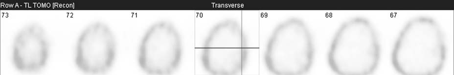
Technique
 Low-energy, high-resolution collimators peaked at 80 keV (Fig. 108.2)
 67Ga SPECT imaging of brain on day 4 with same gamma camera
 Medium-energy collimators
 Triple-peaked at 93, 185, and 300 keV (Fig. 108.3)
Image Interpretation
![]()
Stay updated, free articles. Join our Telegram channel


Full access? Get Clinical Tree


