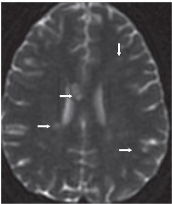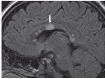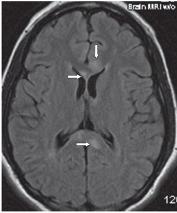


FINDINGS Figures 109-1 and 109-2. Axial DWI with corresponding ADC map through the body of corpus callosum (CC). There are multiple lesions in the CC and white matter (WM) of the corona radiata showing increased diffusion (arrows). Figure 109-3. Sagittal T2 FLAIR. There are multiple CC hyperintense lesions without significant callosal–septal interface signal changes. The large central lesion (arrow) is consistent with the so-called snowball lesion. Figure 109-4. Axial T2 FLAIR through the lateral ventricle. There are multiple hyperintense lesions in the genu and splenium of the CC (arrows).
DIFFERENTIAL DIAGNOSIS Toxic metabolic encephalopathy (medication induced, cocaine, inhaled opiates, chemoradiation, Marchiafava-Bignami), inflammatory demyelination (multiple sclerosis [MS], acute disseminated encephalomyelitis [ADEM], and neuromyelitis optica [NMO]), vasculopathic and vasculitic stroke (venous stroke, systemic lupus erythematosus (SLE), Susac syndrome (SS).
Stay updated, free articles. Join our Telegram channel

Full access? Get Clinical Tree


