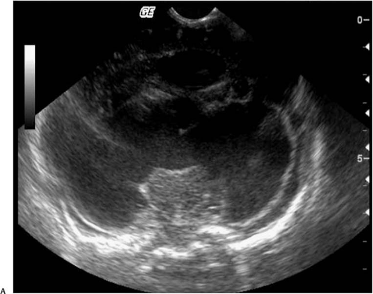
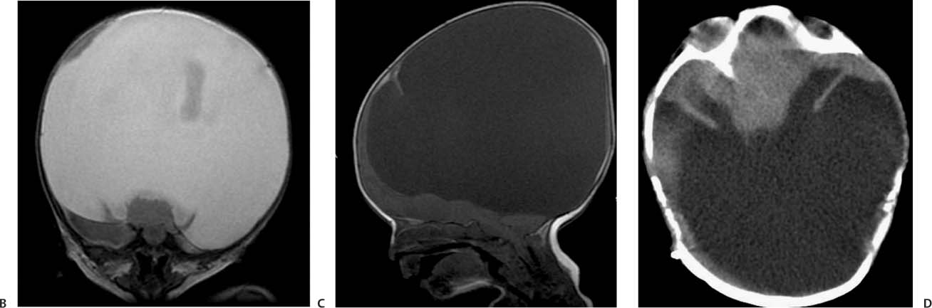
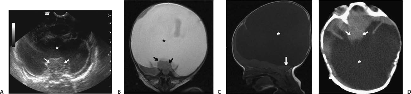
Case 11 A newborn infant with feeding problems and seizures. The baby has a cleft lip. (A) Transfontanellar ultrasound of the brain in a coronal view at the level of the thalami shows a prominent monoventricle (asterisk) and thalamic fusion (arrows). (B) Coronal T2-weighted magnetic resonance image of the brain at the level of the thalami shows a monoventricle, seen as a prominent supratentorial cyst (asterisk), and fusion of the thalami (arrows). Note the absence of a cerebral falx. (C) Sagittal T1-weighted image of the brain shows a large supratentorial cyst (asterisk); note the hypoplasia of the posterior fossa structures (arrow). (D) Axial computed tomography scan of the brain without contrast shows fusion of the thalami (arrows) and a large posterior cyst-monoventricle (asterisk).
Clinical Presentation
Further Work-up
Imaging Findings
Differential Diagnosis
Stay updated, free articles. Join our Telegram channel

Full access? Get Clinical Tree


