 Clinical Presentation
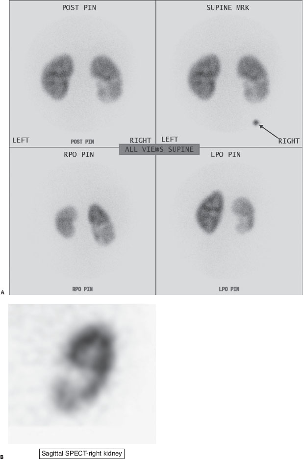
An 8-year-old boy with fever and flank pain.

(A) Tc99m DMSA renal cortical pinhole planar views demonstrate decreased activity to the inferior right kidney (circle). (B) Sagittal SPECT image also shows decreased cortical uptake of the right kidney inferiorly (circle).
 Differential Diagnosis
• Acute pyelonephritis: Regional decreased renal parenchymal uptake makes this the most likely diagnosis.
• Chronic renal scarring: This will also have regional parenchymal defects, but these tend to be more focal and peripheral, and unlike acute pyelonephritis, chronic renal scarring may have associated volume loss.
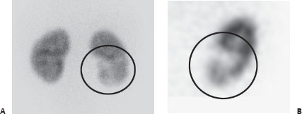
• Renal tumor: Although any space-occupying lesion, including renal cysts or neoplasm, can have decreased uptake, the appearance is typically more focal and masslike, as seen in this DMSA scan from a different patient:


Stay updated, free articles. Join our Telegram channel

Full access? Get Clinical Tree


