 Clinical Presentation
A 43-year-old man with cough and hemoptysis.
 Imaging Findings
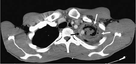
Contrast-enhanced computed tomography (CT): axial image at the level of the thoracic inlet shows a cavitary lesion in the left upper lobe with a crescent-shaped air density (black arrow), peripheral rim enhancement (long white arrow), and a round, denser area of vascular enhancement on its medial aspect (short white arrow).
 Differential Diagnosis
• Rasmussen aneurysm: Infectious aneurysms in a pulmonary artery secondary to pulmonary tuberculosis (TB) are referred to as Rasmussen aneurysms. They characteristically develop as a result of weakening of a pulmonary artery wall adjacent to a TB cavity.
Stay updated, free articles. Join our Telegram channel

Full access? Get Clinical Tree


