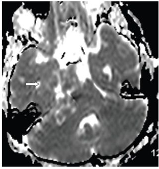
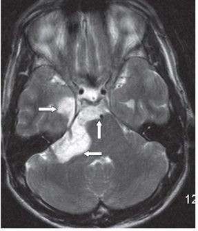
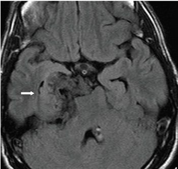
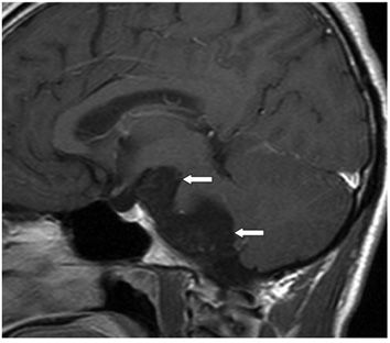
FINDINGS Figures 11-1 and 11-2. Axial DWI with corresponding ADC map through the posterior fossa. There is a heterogeneous DWI hyperintense mass which is isointense (to brain) on ADC map in the right cerebellopontine angle (CPA). The mass extends into the right middle cranial fossa and suprasellar region (arrows). Figure 11-3. Axial T2WI through the posterior fossa. There is a right CPA, prepontine cistern, suprasellar and adjacent right mesiotemporal hyperintense mass with mass effect on surrounding structures (arrows). The intensity is that of CSF. Figure 11-4. Axial FLAIR through the suprasellar cistern. There is a right mesiotemporal/prepontine/upper right CPA somewhat heterogeneous hypointense mass. There is deformity of and mass effect on the brainstem and right temporal lobe with compression of the right temporal horn (arrow). Figure 11-5. Right parasagittal post-contrast T1WI. There is a non-contrast-enhancing lobulated hypointense right CPA and suprasellar mass (arrows) compressing the right cerebellum, brachium pontis, and midbrain.
DIFFERENTIAL DIAGNOSIS Arachnoid cyst, epidermoid cyst, dermoid cyst.
DIAGNOSIS Epidermoid cyst.
DISCUSSION
Stay updated, free articles. Join our Telegram channel

Full access? Get Clinical Tree


