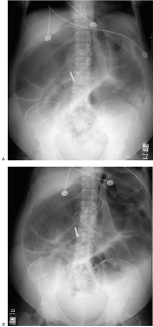
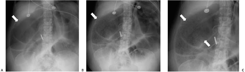
Case 11 A 56-year-old woman presents with marked abdominal distension. Serial radiographs were obtained during her hospital admission. (A) Frontal abdominal radiograph shows marked dilatation of the ascending, transverse, and descending colon, most pronounced in the proximal colon (arrow). There is no air in the rectum, suggesting distal obstruction. (B) A later radiograph shows unchanged colonic dilatation (arrow) and interval development of a mosaiclike mucosal abnormality with polygonal, raised plaques separated by barium. The patient has no allergies. (C) Magnified view of the mucosal abnormality (arrows). •
 Clinical Presentation
 Imaging Findings
 Differential Diagnosis
![]()
Stay updated, free articles. Join our Telegram channel

Full access? Get Clinical Tree


