Case 110
Case History
A 40-year-old woman presents with a palpable breast lump. Mammographically, the lump is associated with abnormal calcifications. Sonographically, the lump corresponds to a hypoechoic ill-defined mass. Biopsy of the lump demonstrates infiltrating ductal carcinoma. Sestamibi study demonstrates abnormal activity in both the breast and the axilla. She is treated with preoperative chemotherapy and the palpable lump disappears. However, the mammographic calcifications remain stable. The sonographic mass shrinks but is still visible. The sestamibi exam shows reduction of breast activity and disappearance of the activity in the axilla.
Physical Examination
• right breast: palpable lump at the 11:00 position
• left breast: normal
Mammogram
Calcifications (Fig. 110–1)
• type: fine linear/branching
• distribution: segmental
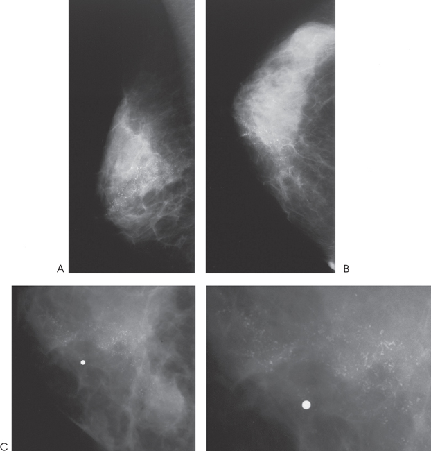
Figure 110–1. In the upper medial breast, there is a segmental group of fine linear branching calcifications. (A). Right ML mammogram. (B). Right CC mammogram. (C). Right CC magnification mammogram.
Ultrasound
Frequency
• 7.5 MHz
Mass
• margin: ill defined
• echogenicity: hypoechoic
• retrotumoral acoustic appearance: increased acoustic transmission
• shape: irregular (Figs. 110–2 and 110–3)


Stay updated, free articles. Join our Telegram channel

Full access? Get Clinical Tree


