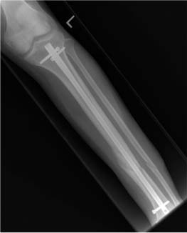
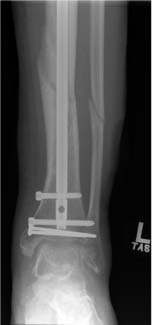
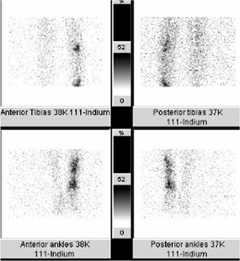
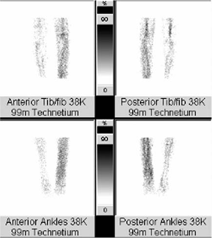
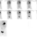
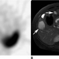
CASE 110 A 38-year-old man with poorly healing fractures of the left tibia that were previously repaired with orthopedic hardware now presents with pain and tenderness in his lower left leg. There is no erythema or draining sinus (Figs. 110.1, 110.2, 110.3, and 110.4). Fig. 110.1 Radiograph of left tibia (knee), anteroposterior projection. Fig. 110.2 Radio-graph of left tibia (ankle), anteroposterior projection. Fig. 110.3 Four views, tibiae (knee to ankle), anterior and posterior projections, 111In-WBCs. Fig. 110.4 Four matching views, tibiae (knee to ankle), anterior and posterior projections, 99mTc-sC. • A 0.5 mCi dose of 111In-WBCs is injected intravenously. • Images 24 hours following tracer injection • Whole-body planar images (not shown) and spot views of the lower legs (Fig. 110.3) obtained with a dual-detector gamma camera • Medium-energy collimators • Energy peaks: 173 and 247 keV • A 12 mCi dose of 99mTc-SC is injected intravenously. • Image at 20 minutes (bone marrow “mapping” scan) • Dual-detector spot views of the lower legs in the same projections used to obtain the WBC spot views (Fig. 110.4)
Clinical Presentation
Technique
111In-WBC Scan
99mTc-SC Scan
Stay updated, free articles. Join our Telegram channel

Full access? Get Clinical Tree


