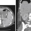
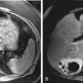
CASE 111
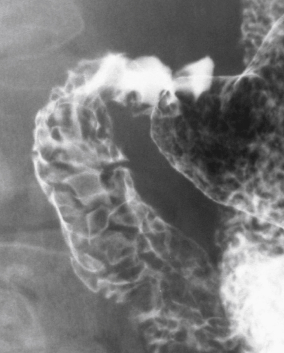
History: A 55-year-old man presents with altered blood in the stool.
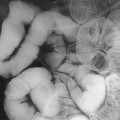
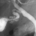
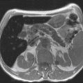
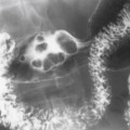
1. What should be included in the differential diagnosis of the imaging finding shown in the figure? (Choose all that apply.)
2. The patient has been undertreated for ulcer disease because he has persistent Helicobacter pylori infection. Which of the following conditions is associated with the highest prevalence of H. pylori infection?
3. What is the most common complication of chronic duodenal ulceration?
4. Which of the following statements regarding the imaging of complications of duodenal ulceration is true?
A. On plain radiography, the most common appearance is gastric outlet obstruction.
Stay updated, free articles. Join our Telegram channel

Full access? Get Clinical Tree


