Case 111
Case History
A 68-year-old woman has increasing calcifications on her screening mammogram. She has past history of lung cancer and bladder cancer. Stereotactic core needle biopsy of the calcifications was not conclusive so a needle localization and excisional biopsy were performed. Patient was unable to maintain an upright position for the needle localization, so sonographic guidance of the wire localization was performed.
Physical Examination
• normal exam
Mammogram
Calcifications (Figs. 111–1 and 111–2)
• type: fine linear/branching
• distribution: linear
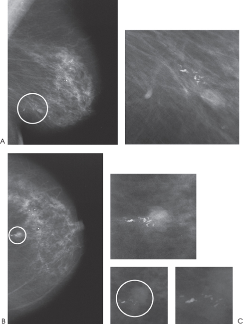
Figure 111–1. In the 6:00 position of the left breast, there is a nodular density associated with linear calcifications (circled). (A). Left MLO mammogram. (B). Left CC mammogram. (C). Left CC magnification mammogram.


Stay updated, free articles. Join our Telegram channel

Full access? Get Clinical Tree


