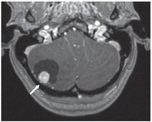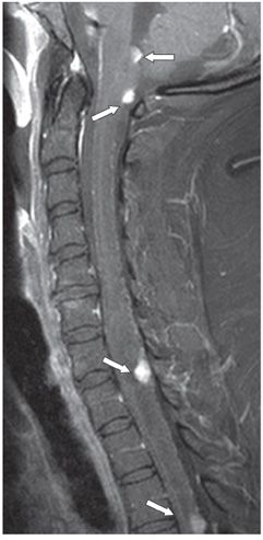

FINDINGS Figure 111-1. Axial T2WI through the posterior fossa. There is a mixed cystic and solid tumor in the right cerebellar hemisphere. A solid nodule (arrow) abuts the pial surface of the cerebellum. Figure 111-2. Axial post-contrast T1WI through the mass. The solid nodule (arrow) demonstrates intense enhancement, but there is a lack of enhancement of the cyst wall. Figure 111-3. Sagittal post-contrast T1WI in a companion case demonstrates multiple hemangioblastomas (arrows) in the posterior fossa, cervical and thoracic spine, diagnostic of von Hippel-Lindau syndrome.
DIFFERENTIAL DIAGNOSIS Hemangioblastoma, pilocytic astrocytoma, glioblastoma, metastasis.
DIAGNOSIS Hemangioblastoma.
DISCUSSION
Stay updated, free articles. Join our Telegram channel

Full access? Get Clinical Tree


