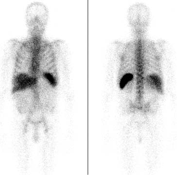
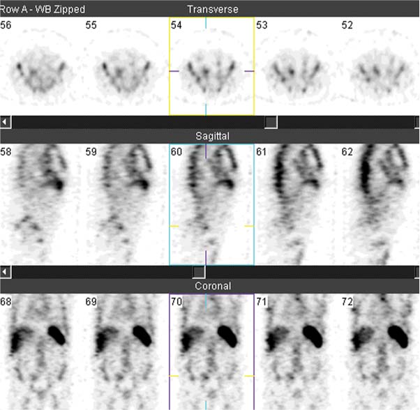
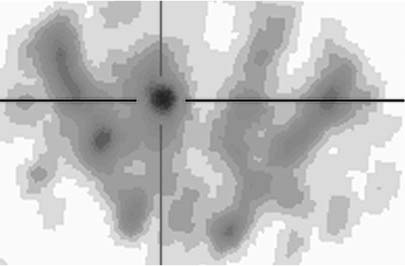
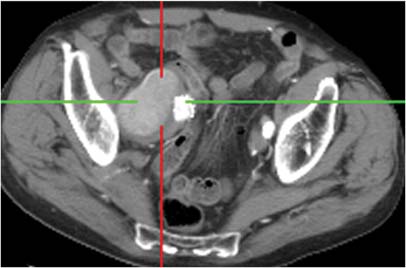
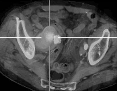
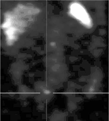
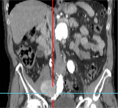
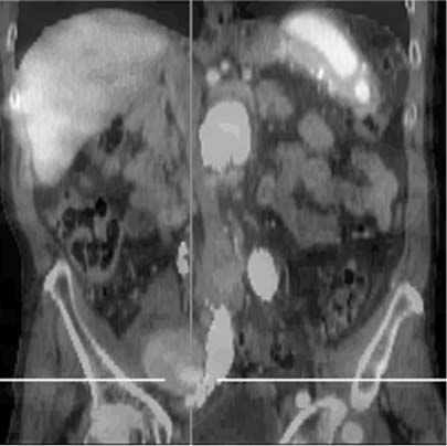
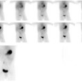
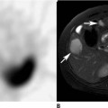
CASE 111 A 72-year-old man develops fever and leukocytosis several weeks after placement of an aortoiliac arterial endograft. Fig. 111.1 Whole-body planar image, anterior and posterior projections, 111In-WBCs. Fig. 111.2 Whole-body SPECT, coronal projection, 111In-WBCs. Fig. 111.3 abdominal SPECT, axial projection, 111InWBCs. Fig. 111.4 abdominal CT, axial projection. Fig. 111.5 Fused SPECT/CT (Figs. 111.3 and 111.4), axial projection. Fig. 111.6 abdominal SPECT, coronal projection, 111In-WBCs. Fig. 111.7 abdominal CT, coronal projection. Fig. 111.8 Fused SPECT/CT (Figs. 111.6 and 111.7), coronal projection. • A 0.5 mCi dose of white blood cells labeled with 111In-WBCs is injected intravenously. • Images are obtained 24 hours following tracer injection. • Whole-body planar imaging (Fig. 111.1) and whole-body SPECT imaging (Fig. 111.2) with a dual-detector gamma camera • Medium-energy collimators • Energy peaks: 173 and 247 keV • Use of software to fuse SPECT and CT images in axial and coronal projections (Figs. 111.3, 111.4, 111.5, 111.6,
Clinical Presentation
Technique
![]()
Stay updated, free articles. Join our Telegram channel

Full access? Get Clinical Tree


