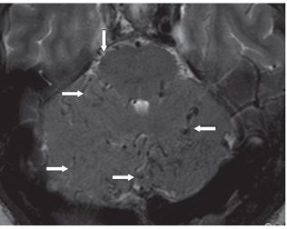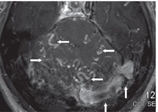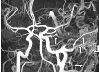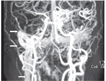


FINDINGS Figure 112-1. Axial T2 FLAIR through the trigones. There are multiple serpentine signal voids (arrows) in the left occipital lobe subarachnoid spaces. Figure 112-2. Axial T2WI through the lower pons. There are multiple serpentine signal voids within the subarachnoid spaces (arrows). Figure 112-3. Axial post-contrast T1WI through the lower pons. There are multiple contrast-enhancing leptomeningeal vessels in the posterior fossa (arrows). The left transverse sinus is enlarged and enhanced (vertical arrows). Figure 112-4. 3D TOF MRA of the head. There are multiple arterial branches (arrows) from the left occipital artery, left middle meningeal artery, and a left internal carotid artery (ICA) tentorial artery feeding a left occipital fistulae in the left transverse sigmoid junction (double arrows). There are multiple draining veins. Figure 112-5. Coronal MIP contrast-enhanced MRV of the head. The left sigmoid sinus and internal jugular veins are not visualized. The right sigmoid sinus and internal jugular vein (arrows) and the bilateral transverse sinuses are visualized.
DIFFERENTIAL DIAGNOSIS Dural arteriovenous fistula (DAVF), leptomeningeal angiomatosis, arteriovenous malformation (AVM).
Stay updated, free articles. Join our Telegram channel

Full access? Get Clinical Tree


