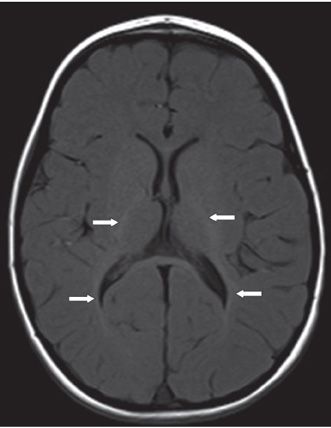
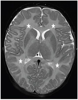
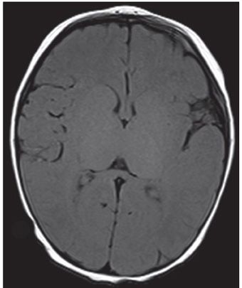
FINDINGS Figure 113-1. Axial T2WI through the basal ganglia. There is diffusely hyperintense white matter (WM) (stars), indicating lack of mature myelin at 1 year of age. Figure 113-2. Corresponding axial T1WI in the same patient. There is normal hyperintense myelin only at the posterior limbs of the internal capsules and along the optic radiations (arrows). Figures 113-3 and 113-4. Axial T2WI with corresponding T1WI through the basal ganglia, respectively, in a companion case. There is diffuse hyperintense myelin on T2WI (stars) and lack of normal hyperintense myelin on T1WI throughout the brain.
DIFFERENTIAL DIAGNOSIS Pelizaeus-Merzbacher disease (PMD), metachromatic leukodystrophy, Lowe syndrome, Salla disease, trichothiodystrophy.
DIAGNOSIS PMD.
DISCUSSION


Stay updated, free articles. Join our Telegram channel

Full access? Get Clinical Tree


