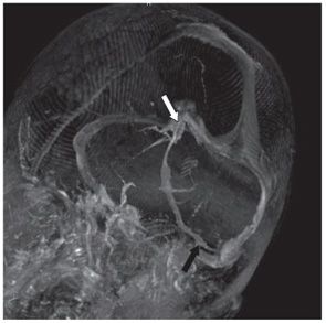
FINDINGS Figure 114-1. Axial post-contrast T1WI through the pons. There is a branching tubular structure emanating from the 4th ventricular region crossing the right middle cerebellar peduncle (arrow). The surrounding brain is normal in appearance. Figure 114-2. 3D volume-rendered MRV, viewing the posterior fossa from a bottom-left vantage, shows the full extent of the lesion, as it originates in the right cerebellum (black arrow), coursing medially and superiorly to join the deep venous system at the vein of Galen (white arrow).
DIFFERENTIAL DIAGNOSIS Developmental venous anomaly (DVA), arteriovenous malformation (AVM), capillary telangiectasia.
Stay updated, free articles. Join our Telegram channel

Full access? Get Clinical Tree


