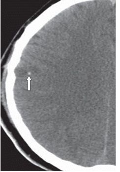
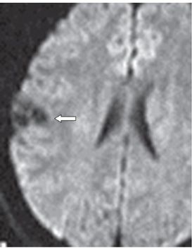
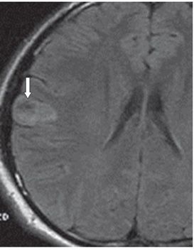
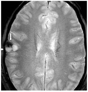
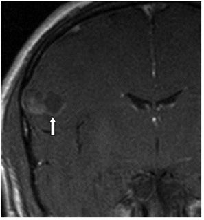
FINDINGS Figures 115-1 and 115-2. Axial contiguous NCCT through the level of the corona radiata. There is a cortical-based suprasylvian right frontal lobe mass with a hyperdense lateral component and a hypodense medial component. There is scalloping of the adjacent inner table (arrow in Figure 115-1) and a medial peripheral focal calcification (arrow in Figure 115-2). Figure 115-3. Axial DWI through the mass. There are three areas of increased diffusion within the mass. Figure 115-4. Axial T2 FLAIR. The mass is well circumscribed and hyperintense with a small area of profound hypointensity anterolaterally (arrow). There is no surrounding edema. Figure 115-5. Axial GRE. There is an area of blooming anterolaterally corresponding to the hypointensity on FLAIR and hyperdensity on CT most probably a focus of hemorrhage or calcification (vertical arrow). The scalloping of the inner table is again demonstrated (transverse arrow). Figure 115-6


Stay updated, free articles. Join our Telegram channel

Full access? Get Clinical Tree


