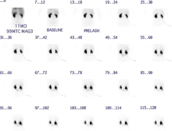
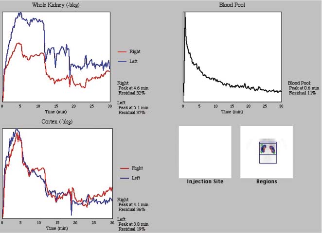
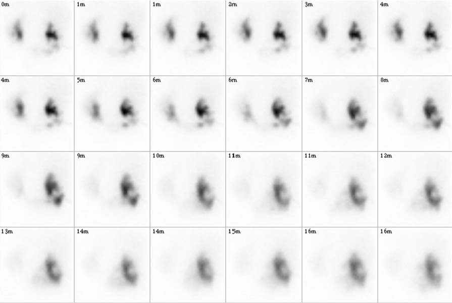
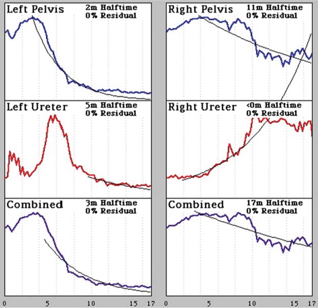
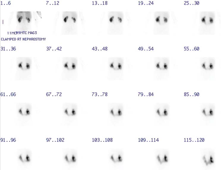
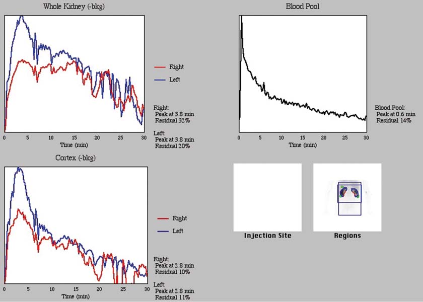
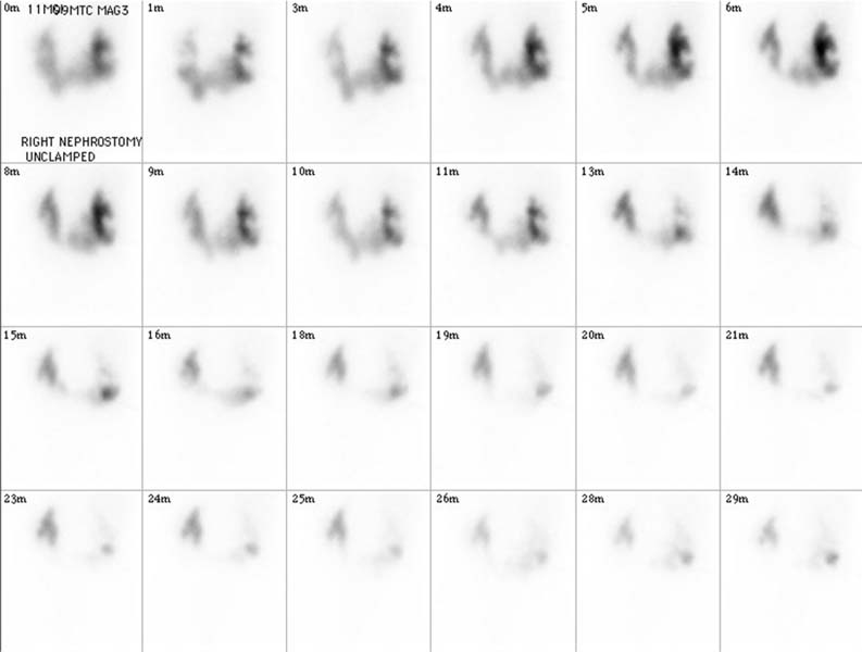
CASE 115 A 39-year-old woman with a history of congenital bladder exstrophy, repaired with an ileal loop and Indiana pouch, presents with recurrent pyelonephritis and severe right-sided flank pain. Fig. 115.1 Fig. 115.2 Fig. 115.3 Fig. 115.4 Fig. 115.5 Fig. 115.6 Fig. 115.7 Fig. 115.8 • Inject 5–10 mCi of 99mTc-MAG-3 intravenously. • Administer 20 mg of furosemide intravenously at 20 minutes after tracer injection. • Use a low-energy, all-purpose collimator. • Energy window 20% centered at 140 keV. • Imaging time is a dynamic sequence: 24 frames at 2 seconds per frame, 16 frames at 15 seconds per frame, and 60 frames at 30 seconds per frame. The patient had a right nephrostomy tube. Dynamic images (Fig. 115.1) obtained in the posterior position with the tube unclamped show that excretion from the right kidney is primarily through the tube. The time–activity curves (Fig. 115.2) show a split function: left 56% and right 44%. Tracer accumulation was seen in the right renal pelvis; however, this cleared substantially following the administration of 40 mg of furosemide (Figs. 115.3 and 115.4).
Clinical Presentation
Technique
Image Interpretation
Stay updated, free articles. Join our Telegram channel

Full access? Get Clinical Tree


