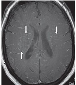
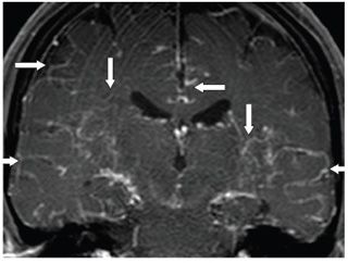
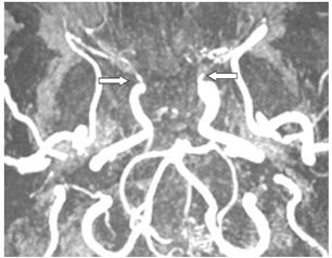
FINDINGS Figure 116-1. Axial T2WI through the suprasellar cistern. There is extensive a network of serpentine signal voids within the suprasellar and perimesencephalic cisterns (arrows) consistent with multiple collaterals. The anterior cerebral artery (ACA) and middle cerebral artery (MCA) are not visualized. Figures 116-2 and 116-3. Axial and coronal post-contrast T1WI through the lateral ventricles, respectively. There is extensive network of contrast enhancement within the subarachnoid spaces (transverse arrows) and transmedullary (vertical arrows). Figure 116-4. 3D TOF MRA. There is bilateral supraclinoid internal carotid artery (ICA) occlusion at the level of the ophthalmic arteries (arrow). The ACA and MCA are not seen bilaterally. There is preservation of the posterior circulation.
DIFFERENTIAL DIAGNOSIS N/A.
DIAGNOSIS Moyamoya disease (MMD).


Stay updated, free articles. Join our Telegram channel

Full access? Get Clinical Tree


