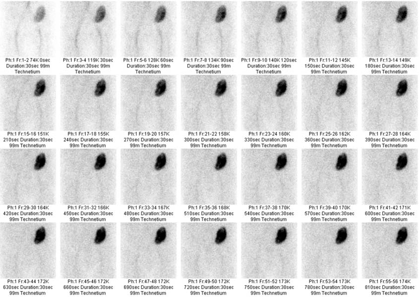
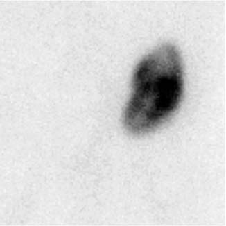
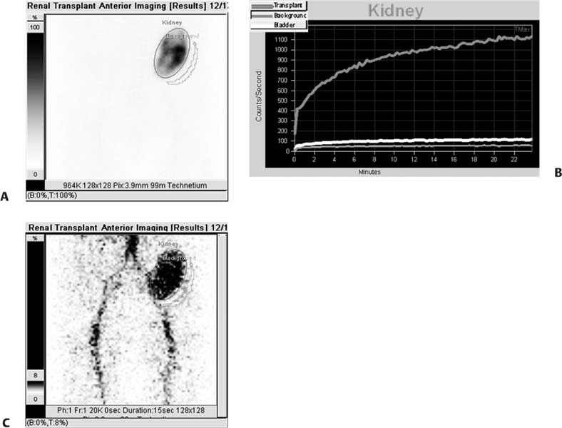
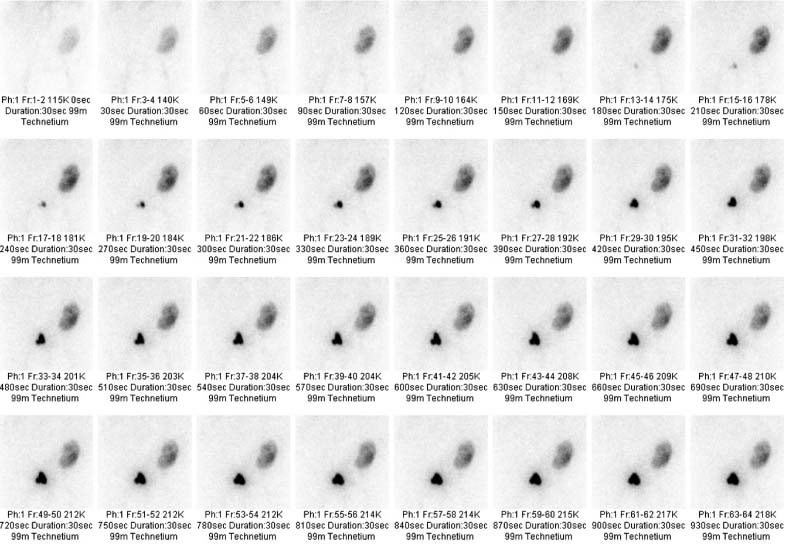
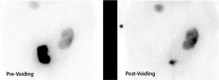
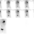
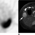
CASE 116 A 54-year-old woman is anuric for 2 days after receiving a renal transplant. Fig. 116.1 Two days post-transplant. Fig. 116.2 Two days post-transplant. Fig. 116.3 Two days post-transplant. Fig. 116.4 Three weeks post-transplant. Fig. 116.5 Three weeks post-transplant. • Inject 10 mCi of 99mTc-MAG-3 with a tight intravenous bolus. 99mTc-DTPA (10–20mCi) can also be used. • Use a low-energy, medium-resolution collimator. • Image native kidneys in the posterior view and pelvic transplant kidneys in the anterior view. • Image for the first minute with 1- to 2-second frames. Then image for 2 to 30 minutes with 30- to 60-second frames. A 5-minute static image at the end may help to demonstrate ureteral uptake. • A 1mg/kg (max 40 mg) dose of furosemide can be administered if the initial set of images suggests obstruction. Image another 20 to 30 minutes after the furosemide injection, and compute the final residual activity in the collecting system (renal pelvis ± ureter) plus the half-time of emptying (see Case 115). Anterior dynamic images (Fig. 116.1) obtained on postoperative day 2 show prompt perfusion of the transplant kidney in the left iliac fossa. This key finding excludes arterial thrombosis as a cause of the patient’s anuria. There is good cortical uptake, with at most a slight lack of homogeneity, and blood pool activity visibly decreases as tracer is concentrated in the kidney. However, no activity is seen in the renal pelvis, ureter, or bladder, either on the dynamic images or on a 5-minute static image taken after 30 minutes (Fig. 116.2). The time–activity curve (Fig. 116.3) shows steadily increasing uptake, which does not maximize until the end of the study. Anterior dynamic images taken from a repeated study 3 weeks later (Fig. 116.4
Clinical Presentation
Technique
Image Interpretation
![]()
Stay updated, free articles. Join our Telegram channel

Full access? Get Clinical Tree


