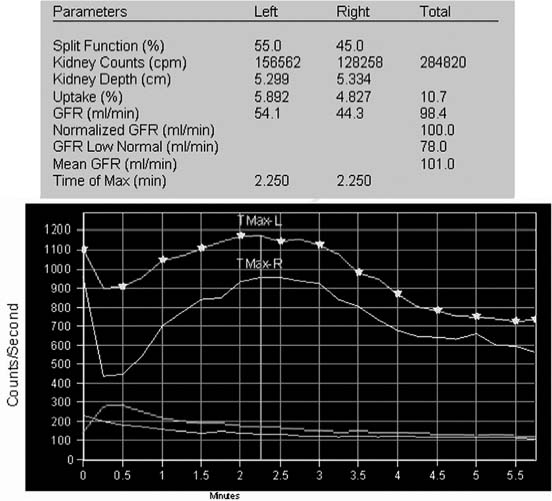
CASE 117 A 48-year-old woman in generally good health presents for evaluation as a prospective renal transplant donor. Fig. 117.1 Fig. 117.2 • Best results are obtained with a medium-energy collimator, although a low-energy, all-purpose collimator can also be used. • Administer 15 mCi of 99mTc-DTPA in a compact bolus injection. • Obtain initial and post-injection syringe counts for 1 minute. • Acquire twenty-four 15-second frames using a 128×128 matrix. Draw regions-of-interest for the whole kidney, cortex, and background. Posterior dynamic images (Fig. 117.1) fail to show the aortic tracer bolus, indicating that imaging was started several seconds late. However, some cardiac and aortic blood pool activity is still seen in the first frame. The venous phase of blood pool activity persists for 2 to 3 minutes in the liver and spleen. This overlaps the renal regions-of-interest and adds to the measured counts in the earliest phase of the study. Both kidneys appear similar in size, with homogeneous cortical uptake. After a cortical transit time of about 2 minutes, tracer can be seen in the collecting systems of both kidneys. At the end of 6.5 minutes of imaging, nearly half of the maximal tracer activity has cleared from both renal cortices. The shape of the time–activity curve differs from those of the other cases in this section because of the shorter imaging time: 6 minutes versus 20 or 30 minutes (Fig. 117.2). There is a transient, steeply dropping peak during the first 15 seconds of the study, which is due to the superimposed blood pool activity from the liver and spleen. • Normal glomerular filtration rate (GFR) Normal renal function, with a normalized total GFR of 100 mL/min per 1.73 m2. The serum creatinine at the time of the study was 0.74 mg/dL. The estimated GFR, based on this serum creatinine level, was calculated to be 84 mL/min per 1.73 m2. Abdominal CT showed normal kidneys bilaterally. No further clinical evaluation was performed. To date, no renal transplant has been performed.
Clinical Presentation
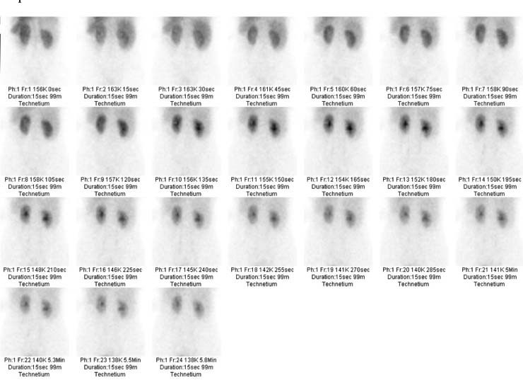
Technique
Image Interpretation
Differential Diagnosis
Diagnosis and Clinical Follow-Up
Discussion
Related posts:


Stay updated, free articles. Join our Telegram channel

Full access? Get Clinical Tree


