Case 118
Case History
A 36-year-old woman felt two left breast lumps. Histology from biopsy of both lumps was infiltrating ductal carcinoma. The patient’s mammograms were interpreted as normal and the surgeon recommended a lumpectomy. The patient was seen at our institution for a second opinion.
Physical Examination
• left breast: two healing biopsy incisions at the 1:30 position about 5 cm apart; no other palpable masses
• right breast: normal exam
Mammogram
• asymmetric density (Fig. 118–1)
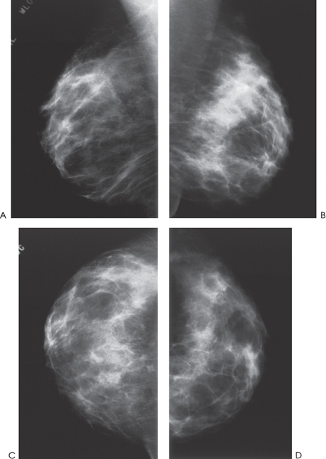
Figure 118–1. There is a band of asymmetric increased density in the posterior third of the left breast on the MLO view. This asymmetry is not evident on the left CC view. (A). Right MLO mammogram. (B). Left MLO mammogram. (C). Right CC mammogram. (D). Left CC mammogram.
Ultrasound
Frequency
• 10 MHz
Mass
• margin: ill defined
• echogenicity: hypoechoic
• retrotumoral acoustic appearance: bilateral edge shadowing


Stay updated, free articles. Join our Telegram channel

Full access? Get Clinical Tree


