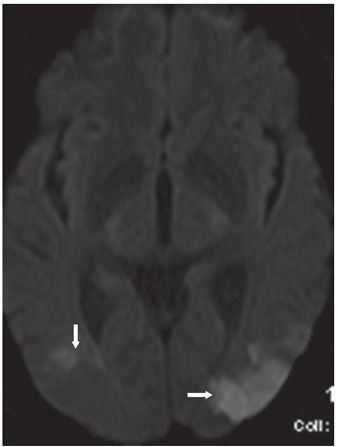
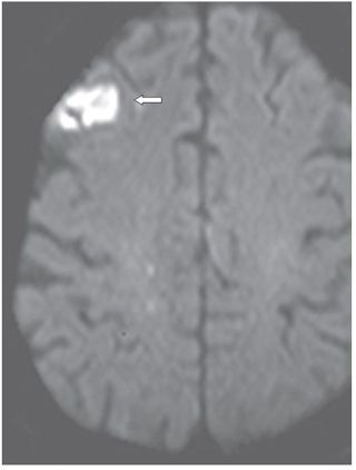
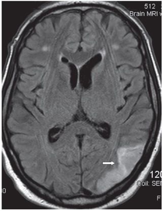
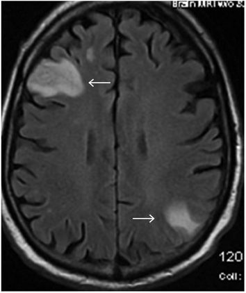
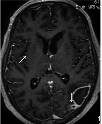
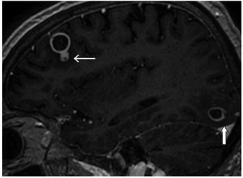
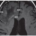
FINDINGS Figures 118-1 to 118-3. Representative axial DWI through various levels of the brain. Figure 118-1 shows multifocal small right cerebellar restricted diffusion (arrow). Figure 118-2 shows a right peritrigonal (vertical arrow) and a left parieto-occipital junction (transverse arrow) areas of restricted diffusion. Figure 118-3 shows a small right frontal cortical/subcortical restricted diffusion (arrow). Figure 118-4. Axial FLAIR through trigone shows a left parieto-occipital junction T2 hyperintensity corresponding to the lesion in Figure 118-2. There are many more lesions on DWI and FLAIR and T2WI than presented here. There was no significant contrast enhancement at presentation, but punctate hemorrhages were present. Figure 118-5. Axial FLAIR through the centrum semiovale at 3 weeks follow-up. There is progression of a right frontal lobe and a left parietal lobe cortical/subcortical hyperintensities (arrows) consistent with infarcts. Figures 118-6 and 118-7


Stay updated, free articles. Join our Telegram channel

Full access? Get Clinical Tree


