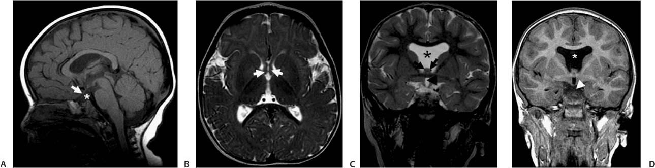
Case 12 A 20-year-old man with a history of bilaterally decreased eyesight and hypopituitarism. (A) A T1-weighted sagittal image of the brain shows a small optic chiasm (arrow) and absence of the pituitary stalk (asterisk). (B) A T2-weighted axial image of the brain shows two separate anterior horns of the fornix (arrows). (C) A T2-weighted sagittal image of the brain shows absence of the septum pellucidum (asterisk). Note the lack of fusion of the fornix (arrows) and the hypoplastic sella, with no evidence of a pituitary gland or pituitary stalk. Hypoplasia of the optic nerves is seen (arrowhead). (D) A T2-weighted sagittal image of the brain shows absence of the septum pellucidum (asterisk). Note the hypoplastic sella, with no evidence of a pituitary gland or pituitary stalk. Hypoplasia of the optic chiasm is seen (arrowhead).
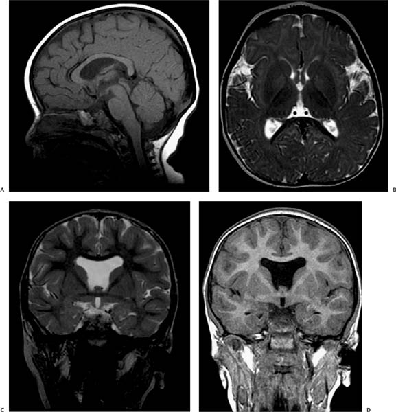
Clinical Presentation
Imaging Findings
Stay updated, free articles. Join our Telegram channel

Full access? Get Clinical Tree


