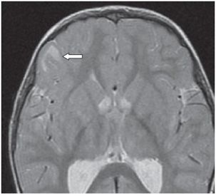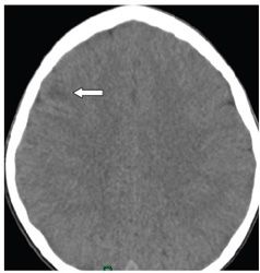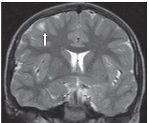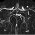


FINDINGS Figures 12-1 and 12-2. Axial T2WI and GRE through the frontal lobes demonstrate expanded right frontal gyrus with ill-defined hyperintensity in the subcortical white matter (WM) extending in a flame-shaped fashion into the deep WM in Figure 12-1 (arrows). Figure 12-3 and 12-4. Axial NCCT and corresponding coronal T2WI through the frontal lobes in a companion case demonstrate a right frontal lobe subcortical hypodensity on CT and corresponding subcortical hyperintensity on the coronal T2WI (arrows).
DIFFERENTIAL DIAGNOSIS Gliosis, polymicrogyria, tuberous sclerosis, low-grade tumor, focal cortical dysplasia.
DIAGNOSIS Focal cortical dysplasia (balloon cell focal cortical dysplasia of Taylor [FCDT]).
DISCUSSION
Stay updated, free articles. Join our Telegram channel

Full access? Get Clinical Tree


