Case 12
 Clinical Presentation
A 24-year-old woman with lower abdominal pain, fever, and leukocytosis.
 Imaging Findings
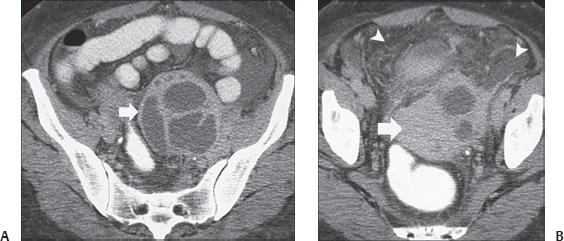
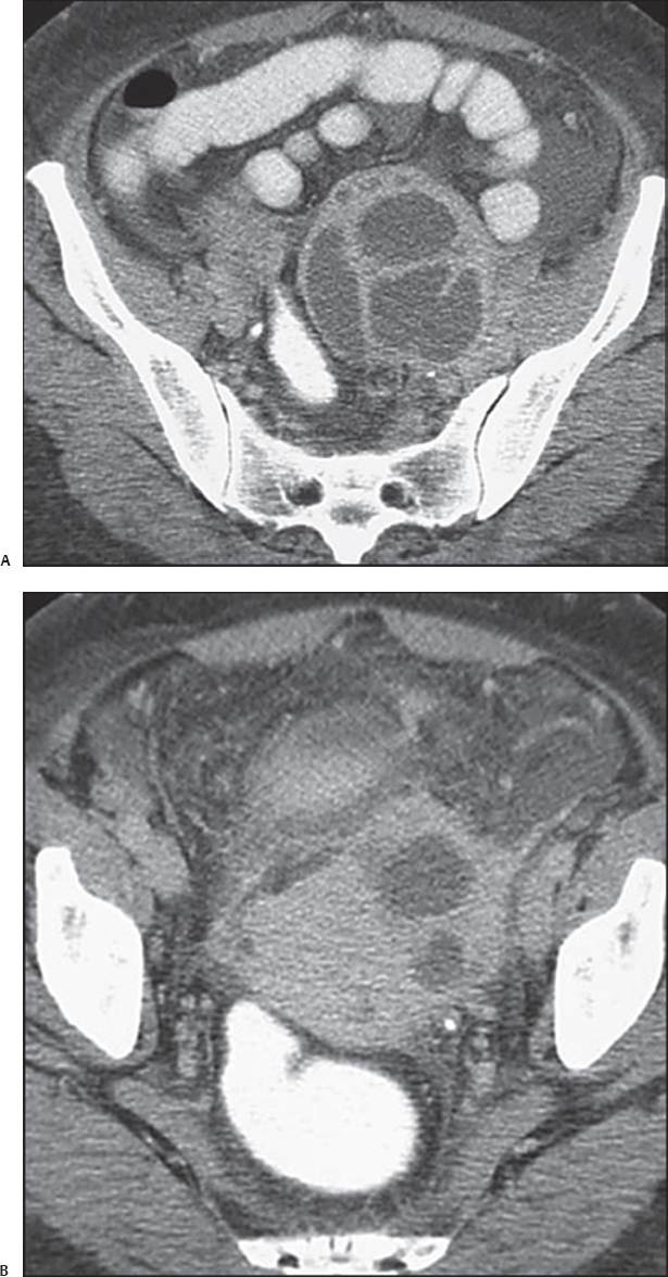
(A) Contrast-enhanced computed tomography (CT) image of the pelvis shows a large, multilocular mass (arrow) on the left. The mass contains fluid. The walls and septa are thick but regular and show some enhancement. (B) Contrast-enhanced CT image of the pelvis at a level inferior to that of Figure A shows the uterus (arrow) displaced to the right by the mass, which is therefore in the adnexa. Stranding and fluid are seen in the pelvic peritoneal fat (arrowheads). The ovaries are not visualized.
 Differential Diagnosis
• Left tubo-ovarian abscesses: Adnexal fluid collections that are unilateral or bilateral and unilocular or multilocular, with enhancing walls and surrounding peritoneal stranding and fluid, in a patient who has clinical signs of sepsis are characteristic of tubo-ovarian abscesses.
• Ovarian neoplasm:
Stay updated, free articles. Join our Telegram channel

Full access? Get Clinical Tree


