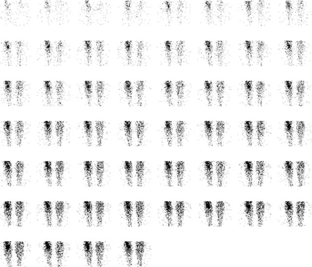
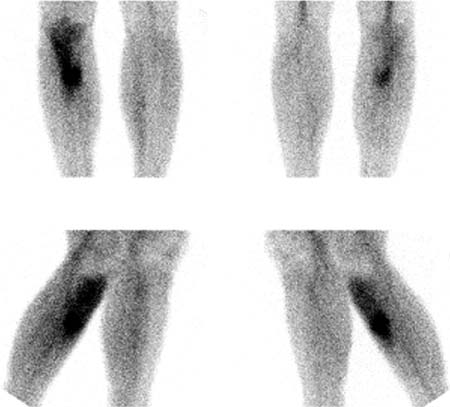
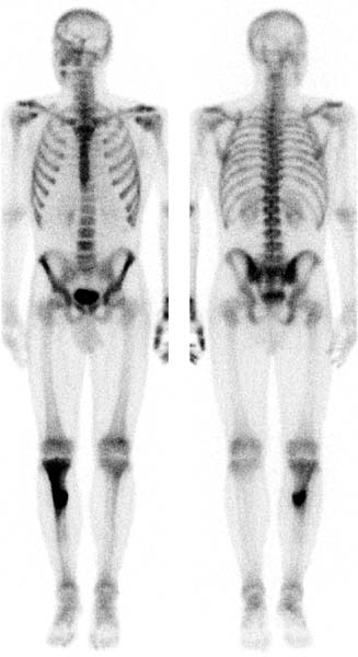
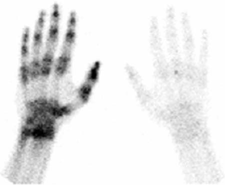
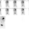
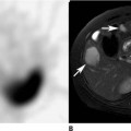
CASE 12 A 22-year-old baseball pitcher presents with right shin pain (Figs. 12.1, 12.2, and 12.3). Fig. 12.1 Fig. 12.2 Fig. 12.3 Fig. 12.4 • A 20 mCi dose of 99mTc-MDP is administered intravenously. • Flow images of the pelvis are obtained for 1 second per frame at the time of tracer injection in the anterior and posterior projections. (For display purposes, images are summed at 3 seconds per frame.) • A blood pool image is obtained for 3 minutes within 10 minutes of tracer injection. • Whole-body images of the skeleton are obtained 3 hours after tracer administration. • A 1024 × 256 matrix is used for whole-body images. • Emphasize the importance of oral hydration to improve soft tissue and bladder clearance. Blood flow images (Fig. 12.1) show increased flow to the right proximal tibia. Blood pool images (Fig. 12.2) in multiple projections show increased tracer localization extending from the right tibial plateau into the proximal one-third of the diaphysis of the tibia. The uptake is predominantly in the anterior aspect of the right tibia. Delayed whole-body images in the anterior and posterior projections (Fig. 12.3) show findings similar to those in the blood pool images; however, additional abnormal uptake is shown within the left hand and wrist.
Clinical Presentation
Technique
Image Interpretation
Stay updated, free articles. Join our Telegram channel

Full access? Get Clinical Tree


