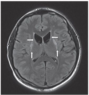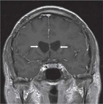

FINDINGS Figure 120-1. Axial T2WI through the caudate nucleus. There is outward bowing of the lateral walls of the frontal horns with nonvisualization of the heads of the caudate nuclei (arrows). There is hyperintensity of the putamina (vertical arrows). Figure 120-2. Corresponding FLAIR image shows similar findings (arrows). Figure 120-3. Coronal post-contrast T1WI shows the “box-like” configuration of the frontal horns of the lateral ventricles (arrows). There is no contrast enhancement.
DIFFERENTIAL DIAGNOSIS Huntington disease, neuroacanthocytosis syndromes (McLeod, chorea-acanthocytosis), frontotemporal dementia, and Alzheimer disease.
DIAGNOSIS Huntington disease.
DISCUSSION MRI shows characteristic atrophy involving the head of the caudate nucleus and the putamen. Caudate atrophy results in the loss of the bulge of the inferior lateral borders of the frontal horns with enlargement of frontal horns of the lateral ventricles and flattening of their lateral contour giving them a “box-like” configuration. The brain is diffusely atrophic with white matter (WM) volume loss affecting both frontal lobes. DWI reveals increased ADC in the caudate nucleus, putamen, and WM which correlates with disease severity. Signal intensity abnormalities consisting of slight T2 hyperintensity in the atrophic neostriatum correlates with greater motor and cognitive impairments and usually seen in the juvenile form. Proton MRS shows low levels of N
Stay updated, free articles. Join our Telegram channel

Full access? Get Clinical Tree


