Case 121
Case History
A 67-year-old female, transferred from another medical facility, was evaluated for abdominal pain. Abdominal computed tomography (CT) demonstrated multiple low-density liver masses. Liver biopsy indicated metastatic adenocarcinoma.
Physical Examination
• normal exam
Mammogram
• focal asymmetric density (Fig. 121–1)
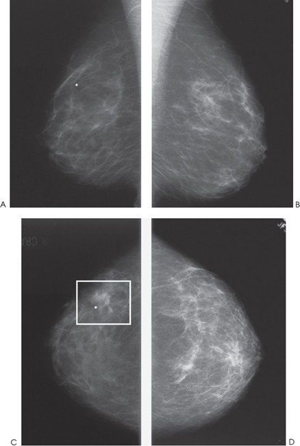
Figure 121–1. A focal asymmetric density (square) in the right outer breast is visible only on the CC view. This density has been unchanged for 5 years. (Radiopaque marker has been placed on skin to aid locating the density on other views.) (A). Right MLO mammogram. (B). Left MLO mammogram. (C). Right CC mammogram. (D). Left CC mammogram.
Ultrasound
Low Frequency
Frequency
• 7 MHz
Mass
• margin: ill defined
• echogenicity: hypoechoic


Stay updated, free articles. Join our Telegram channel

Full access? Get Clinical Tree


