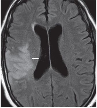
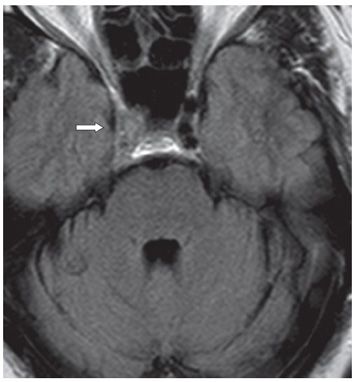
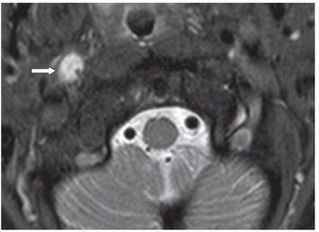
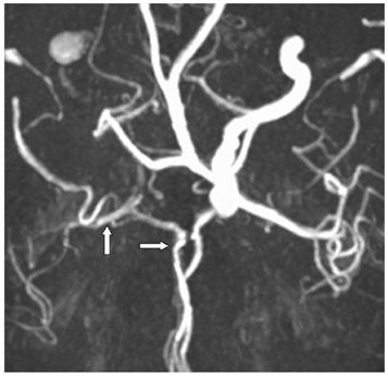
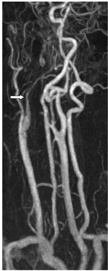
FINDINGS Figure 121-1. Axial DWI through the lateral ventricles. There is a large cortical subcortical posterior right frontal hyperintensity (arrow), which extends into the right parietal lobe on other slices. This region has low ADC (not shown) consistent with acute/subacute ischemic infarct in the right middle cerebral artery (MCA) inferior division territory. Figure 121-2. Corresponding axial FLAIR. There is a smudgy hyperintensity in the right MCA territory posteriorly (arrow). Figure 121-3. Axial FLAIR through the cavernous sinuses. The usual signal void in the right internal carotid artery (ICA) is absent replaced by isointensity (arrow) consistent with occlusion of the right ICA. Figure 121-4
Stay updated, free articles. Join our Telegram channel

Full access? Get Clinical Tree


