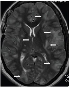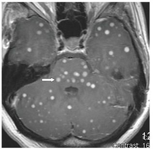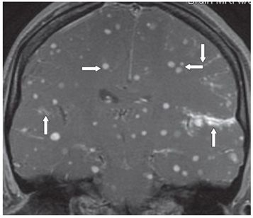


FINDINGS Figures 124-1 and 124-2. Axial FLAIR through the posterior fossa and T2-weighted MRI through the level of the basal ganglia. There are multifocal smudgy cortical/subcortical hyperintensity widespread in the brainstem, cerebellum, and bilateral cerebral hemispheres (arrows point to representative lesions) with some central gray matter components with features consistent with vasogenic edema. Figures 124-3 and 124-4. Axial post-contrast T1WI through the posterior fossa and coronal post-contrast T1WI through the sylvian fissures. There are numerous tiny nodular contrast-enhancing lesions throughout the brainstem, cerebellum, and cerebral hemispheres mainly in cortical and subcortical locations (arrows point to representative lesions). Multiple areas of leptomeningial enhancement are also demonstrated in Figure 124-4 (vertical arrows).
DIFFERENTIAL DIAGNOSIS Miliary tuberculosis (TB), metastases, neurocysticercosis, fungal granulomas.
DIAGNOSIS Miliary TB of the brain.
DISCUSSION
Stay updated, free articles. Join our Telegram channel

Full access? Get Clinical Tree


