Case 125
Case History
An 81-year-old woman presents for screening mammogram.
Physical Examination
• normal exam
Mammogram
• architectural distortion (Fig. 125–1)
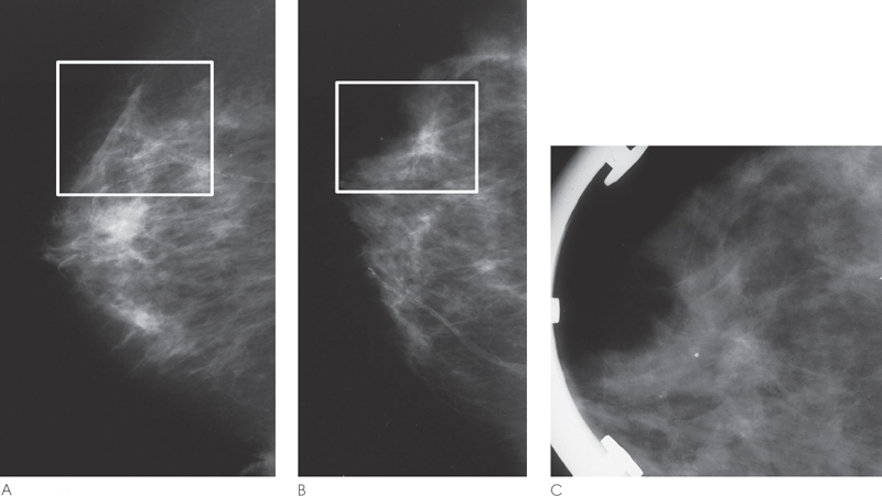
Figure 125–1. In the right upper outer quadrant there is peripheral architectural distortion (outlined by squares). In the ML view, the superior parenchymal contour is abnormally triangular (A) and in the CC view the anterior parenchymal line is retracted (B). (A). Right ML mammogram. (B). Right CC mammogram. (C). Right CC spot compression.
Ultrasound
Low Frequency
Frequency
• 8 MHz
Mass
• margin: ill defined
• echogenicity: hypoechoic


Stay updated, free articles. Join our Telegram channel

Full access? Get Clinical Tree


