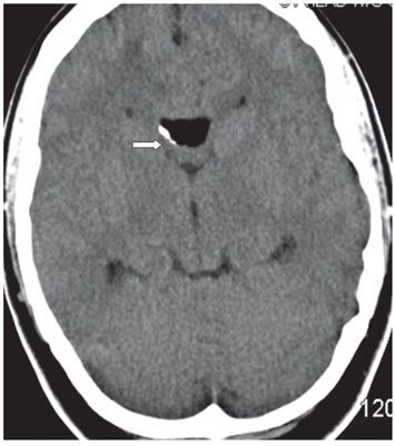
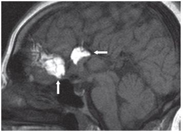
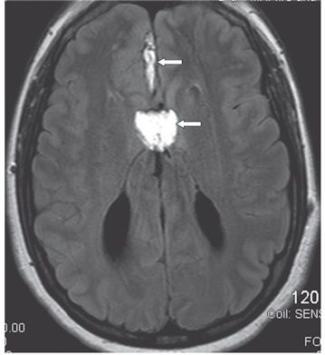
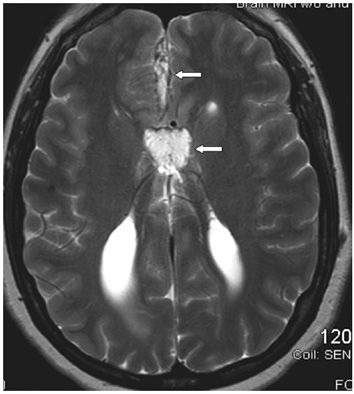
FINDINGS Figures 126-1 and 126-2. Axial NCCT through the suprasellar and third ventricular levels, respectively. There are multiple hypodense masses in the right sylvian fissure, anterior interhemispheric fissure on Figure 126-1 and in the region of the genu of corpus callosum (CC) in Figure 126-2 (arrows) with HU of −105 characteristic of lipoma. There is a linear hyperdensity on the right side of the fat in Figure 126-2 consistent with calcification. Figure 126-3. Sagittal MRI T1WI. There is a large hyperintensity in the anterior interhemispheric fissure (vertical arrow) and at the location of the genu of the CC (transverse arrow). The CC is absent. Figures 126-4 and 126-5. Axial FLAIR and T2WI, respectively, through the level of the lateral ventricles. There is a thick anterior interhemispheric hyperintensity and a triangular-shaped hyperintensity between the tapered and displaced frontal horns of the lateral ventricles in the location of the absent CC (arrows). Figure 126-6


Stay updated, free articles. Join our Telegram channel

Full access? Get Clinical Tree


