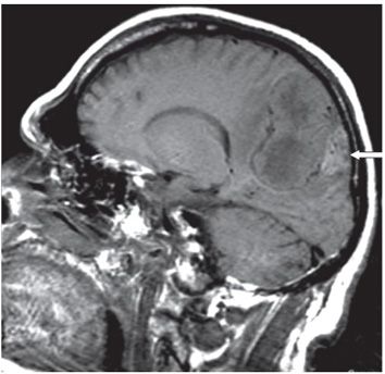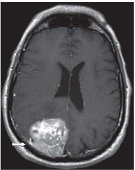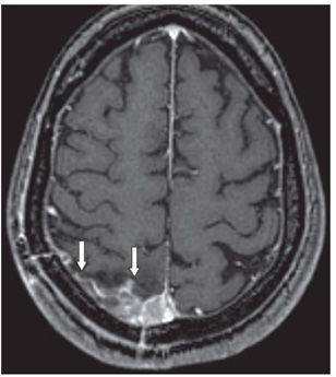


FINDINGS Figure 127-1. Axial T2WI through the parietal lobes. There is a right parietal heterogeneous extraaxial mass posteromedially abutting the falx cerebri and convexity dura. There are prominent flow voids within (vertical arrow). There is surrounding vasogenic edema particularly anteriorly (transverse arrow) with local mass effect. Figure 127-2. Sagittal T1WI through the mass. The mass is heterogeneous with areas of isointensity and hypointensity. Posteriorly, there is T1 hyperintensity suggestive of hemorrhage (arrow). There is no adjacent calvarial hyperostosis. Figure 127-3. Axial post-contrast T1WI. There is heterogeneous but avid enhancement with a small dural tail laterally (arrow). The patient underwent complete resection of the lesion and postoperative baseline study (not shown) revealed no residual abnormal enhancement. Figure 127-4. Axial post-contrast T1WI 3 years later. There is enhancing nodular mass with dural tail within the surgical bed consistent with tumor recurrence (arrows).
DIFFERENTIAL DIAGNOSIS Meningioma, hemangiopericytoma (HPC), dural metastases, lymphoma, neurosarcoidosis, gliosarcoma, solitary fibrous tumor.
DIAGNOSIS Hemangiopericytoma (HPC).
DISCUSSION
Stay updated, free articles. Join our Telegram channel

Full access? Get Clinical Tree


