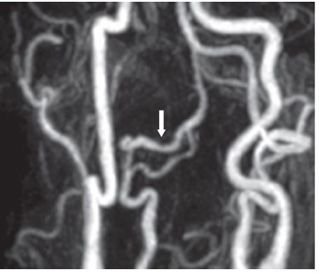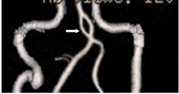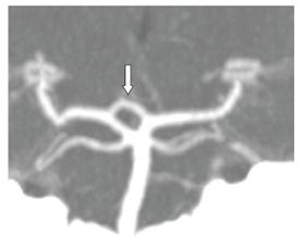


FINDINGS Figure 128-1. 3D TOF MRA volume-rendering submento-vertical (SMV) view. The medial half of the left A1 has two separate segments (arrows). The posterior segment is joined to the anterior communicating artery while the anterior segment is joined to the left A2. The pattern is consistent with fenestration of the left A1. Figure 128-2. Oblique 3D TOF MRA MIP of the neck. There is fenestration of the right distal vertebral artery (arrow) at V3–V4 junction. Figure 128-3. Coronal 3D volume rendering of CTA of the neck. There is fenestration of the proximal basilar artery (arrow). Figure 128-4
Stay updated, free articles. Join our Telegram channel

Full access? Get Clinical Tree


