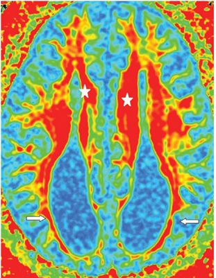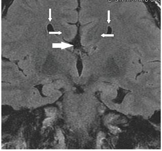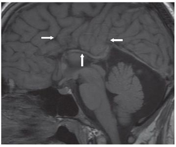


FINDINGS Figure 129-1. Axial T2WI through the lateral ventricles. The lateral ventricles are widely separated and parallel with bilateral tapering of the frontal horns (vertical arrows) and dilated occipital horns (colpocephaly) (transverse arrows). The interhemispheric fissure separates the two lateral ventricles with well-formed subcortical white matter (WM) tract (Probst bundle) and cortical gray matter (GM) separating the lateral ventricles from the interhemispheric fissure. The septum pellucidum and corpus callosum (CC) are absent. Figure 129-2. Axial DTI Fractional Anisotropy (FA) map through the lateral ventricles. The WM tract around the colpocephaly is thin suggesting hypoplasia (arrows). WM tracts do not cross the midline. Probst bundles are medial to the ventricles (star). Figure 129-3. Coronal T2 FLAIR through the frontal horns. There is wide separation of the tapered frontal horns (vertical arrows). WM Probst bundle project medially to the frontal horns (transverse arrows). The interhemispheric fissure extends down to the third ventricular roof (hatched arrow). Figure 129-4. Sagittal T1WI. There is radiating cortical bundles, so-called spoke wheel gyri, in the midline (transverse arrows). The CC and the cingulate gyrus are missing. The internal cerebral vein lies in the inferior aspect of the interhemispheric fissure, the expected location on the velum of the third ventricle (vertical arrow).
Stay updated, free articles. Join our Telegram channel

Full access? Get Clinical Tree


