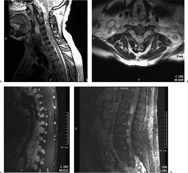
Case 13 A 40-year-old man with progressive weakness of both upper extremities for 1 year. (A) Sagittal T2-weighted image (WI) of the cervical spine demonstrates a widened spinal canal with an intradural, extraspinal mass that compresses the spinal cord posteriorly (arrowhead). A soft-tissue mass in the prevertebral soft tissues shows a high T2 signal (arrow). (B) Axial T2WI of the spine at the level of C5 shows anterior compression of the spinal cord (black arrow) by a hyperintense, lobulated mass that occupies the spinal canal and extends through the neural foramina to the paraspinal soft tissues (arrowheads). Note the widening of the neural foramina (white arrows). (C) Parasagittal fat-saturated T1WI of the thoracic spine with gadolinium shows enhancing masses (arrowheads) at multiple levels that widen the neural foramina and extend to the paraspinal soft tissues (arrows). (D) Sagittal fat-saturated T1WI shows multiple oval, enhancing masses in the spinal canal (arrowheads). • Spinal neurofibromatosis type 1 (NF1; also known as von Recklinghausen disease): Scoliosis is seen in 71% of cases. Plexiform neurofibromas are pathognomonic for NF1; they can have a dumbbell appearance if located in a neural foramen, which they can enlarge. They typically enhance. • Spinal meningioma:
Clinical Presentation
Imaging Findings
Differential Diagnosis
![]()
Stay updated, free articles. Join our Telegram channel

Full access? Get Clinical Tree


